Over 200 modern lizard species possess slits, apertures and folds in the skin that lead to epidermal structures termed mite pockets…

Caption: a mite pocket montage, showing a mite pocket in a scrub lizard (at left), the mite-filled nuchal pockets of a tropidurid (at upper right), and (at lower right) all the locations where pockets can be present. Images: McCoy et al. (2012); Carvalho et al. (2018); Arnold (1986).
Variously located on the sides of the neck, armpit region, and in front of and behind the top of the thigh, these are often inhabited by parasitic mites, specifically chigger mites of the families Trombiculidae and Leeuwenhoekiidae. Because the pockets seemingly don’t have a function other than their use by mites, it was proposed back in the 1980s that lizards and mites have co-evolved such that the pockets are specialized mite aggregation sites.
And because the lizards with these pockets belong to far-flung branches of the squamate family tree (they’re present in various geckos, phrynosomatids, tropidurids, oplurids, chameleons, lacertids and skinks), the pockets must have evolved at least seven times.

Caption: a highly simplified squamate phylogeny (based on recent molecular results) serving to emphasize how pockets (P) have evolved several times in distantly related groups. Even within Iguania (represented here as a single lineage), pockets are present in more than four groups. Image: Darren Naish, produced for my in-prep textbook.
Supporting this co-evolutionary idea is data indicating that the pockets are most prevalent, and largest, in lizards that occupy the same sort of humid, terrestrial habitats as those favoured by mites, and that lizards in arid or arboreal environments generally don’t have them (Arnold 1986, Curtis & Baird 2008, Reed 2014). Also worth noting is that males tend to have higher mite loads than females. I should also add at this point that the mites we’re talking about here are definitely parasitic: none are phoretic mites (those that use larger animals as transport to get from A to B and don’t exact much cost on said larger animal). Scale mites and ticks also use the pockets in some species.

Caption: at left, mites clustered in the right nuchal pocket of a Florida scrub lizard Sceloporus woodi. At right: perhaps casting doubt on the idea that the pockets have co-evolved with mites is the fact that mites cling opportunistically to the skin folds and flaps present in lizards without pockets, like this Agama cristata from Guinea. Images: McCoy et al. (2012); Wagner et al. (2009).
To scientists familiar with lizards (and the literature devoted to them), the existence and claimed function of mite pockets is well known, and indeed the structures have received high-profile coverage in the scientific literature (Benton 1987)… at least, as ‘high profile’ as you can expect for something like this. A memorable back-and-forth in the technical literature also brought the topic to wider attention than it might otherwise have had (Arnold 1986, 1993, Bauer et al. 1990, 1993). And, today, I get the impression from articles online and comments in the literature that the hypothesis that these structures are (1) adaptive and (2) evolved in-step with mite parasitism appears essentially accepted.

Caption: mite pocket distribution as portrayed on an archetypal lizard. Going from head to tail, they are: n = nuchal; a = axillary; pa = post-axillary; i = inguinal; pf = postfemoral. Image: Arnold (1986).
Prior to the appearance of the ‘mite pocket’ idea, these structures were usually given descriptive names related to their anatomical position, such that they were axillary and postfemoral pockets, axillary and inguinal pits, or postfemoral dermal pockets (Arnold 1986, pp. 1-2). The term ‘mite pockets’ was first used by Loveridge (1925) who (after observing them in Asian bent-toed geckos) also suggested that the term acarodomata (singular: acarodomatum) might be used for them*. That technical name wasn’t original to him but came from the botanical literature.
Because, however, calling them ‘mite pockets’ implies that this is certainly what they are, it seems more appropriate to me to be less committal, and for that reason I’m just going to call them ‘pockets’ from hereon.
* The spellings acarodomatia and acarodomatium have also been used (Bertrand & Modrý 2004). Mite-housing structures in hymenopteran insects (yup, they too are a thing) are termed acarinaria, and you might argue that this would be a better term than one devised for plants.

Caption: other organisms have mite-housing structures that have been seen as precedents to the mite pockets of lizards, including plants and insects. These images show the acarinaria of the small Eurasian wasp Allodynerus delphinalis. The wasps transport the mites to their nests where they help protect Allodynerus from attacks by parasitoids. This is therefore a mutualistic relationship. Image: Okabe & Makino (2008).
In memory of Dr Nick Arnold. The seminal work on these structures was published by herpetologist and lizard specialist Nick Arnold in 1986, and here I must make a brief diversion, since Nick died last year and I thought very highly of him, and still do. Dr E. N. Arnold, formally Edwin Nicholas Arnold and based at London’s Natural History Museum where he was Curator of Herpetology, was a brilliant scientist, and he leaves behind an impressive legacy of interesting and compelling studies of lacertid and agamid lizards, recently extinct island-dwelling tortoises and much else. An aspect of Nick’s writing that often stands out is that he was especially good at interpreting or framing data within historical or phylogenetic hypotheses.
Caption: Nick Arnold (1940-2023) was honoured with an obituary in The Times (original is here, but paywalled). Few of us will be considered worthy of being memorialized in this way. Image: (c) The Times.
Many naturalists unfamiliar with the technical literature were also exposed to Nick’s writings, since (with J. A. Burton) he authored the highly successful and very good Collins book A Field Guide to the Reptiles and Amphibians of Britain and Europe, first published in 1978 and illustrated by Denys Ovenden. This was the standard go-to guide to the European herpetofauna for over three decades, and in part still is. Finally: as is true of all the best scientists, Nick was also a phenomenally nice person and was appropriately generous with time and resources when opportunity allowed.

Caption: Nick Arnold’s Collins field guide to the reptiles and amphibians of Europe saw several editions. I don’t have all of them… but I’m working on it. Burton is not credited as a coauthor on the later editions; I don’t know why.
Anyway… back to the pockets. After analysing the anatomy of pockets in geckos, phrynosomatids, chameleons and lacertids, Arnold (1986) concluded that the skin within the pockets was essentially adapted for mite use: the pockets aren’t just epidermal invaginations, they’re invaginations where the skin is distinctly thickened, and where the pocket interior has a modified skin surface, such that scalation is reduced or even absent, and blood supply is rich (Arnold 1986, p. 19). In some pocket-bearing lizards, there are indications that lymphoid cells are densely clustered where mites occur (suggesting a sort of enhanced local immune response to the mites’ mouthparts), but Arnold (1986, p. 17) expressed uncertainty as to whether this was adaptive on the part of the lizards or a consequence of mite activity.

Caption: Arnold (1986) featured several histological investigations of pocket anatomy and what this meant for mite presence. At left, this section through a nuchal pocket on a fence lizard shows how the collagen (c’) within the pocket is thinner than that elsewhere across the body, and how the epidermis around the mite feeding site (the stylostome, marked s) consisted mostly of lymphoid cells (l). At right, this section through a mite and the post-axillary pocket of a Pristurus gecko from Oman shows how the skin in the vicinity of the stylostome (s) is densely packed with lymphoid cells (l). Images: Arnold (1986).
The main take home from Arnold (1986) is that the pockets “reduce the deleterious effects” (p. 18) of parasitic mites by providing mite-friendly areas that cause the mites to cluster in select areas rather than spread across the body, this minimizing tissue damage and irritation more broadly. Arnold’s argument was termed the ‘damage limitation’ or ‘damage-amelioration’ hypothesis by later authors (Bauer et al. 1990, Reed 2014).

Caption: mites clustered in an axillary pocket of a chameleon (Rieppeleon brevicaudatus, though included in Rhampholeon when Arnold was writing). The mites are obviously distinct from the “sculpted surfaces of the scales lining the pockets” and are actually feeding from “the solf folded interstitial skin between the scales” (Arnold 1986, p. 8).
Some quick and dirty speculations. Incidentally, Arnold’s hypothesis that lizards have repeatedly evolved special mite-nurturing pockets raises all sorts of speculative possibilities for lizard-parasite evolution.
If these really are ‘mite pockets’, should we – for example – expect the evolution of dedicated pocket-dwelling parasites of lizards? Think sessile, limbless mites or even pocket-dwelling parasites belonging to other groups that have moved in to take advantage. What about the development of ‘pocket-raiding’ habits by conspecifics or even by other animals? Might it even be that the supposed mite-nurturing anatomy of the pockets could be co-opted to something else, like the feeding of conspecifics? If the pockets really provide enhanced blood flow and have specialized internal skin that’s co-evolved with the feeding on body fluids… well, the parallels with the evolution of lactation in mammals are unavoidable.

Caption: I’m old-fashioned and still rely extensively on physical printed literature. This isn’t necessarily because I want to, but because so much of the material I need to access is still not easily findable in digital form. Here are physical reprints of various of the sacred texts discussed in this article. Image: Darren Naish.
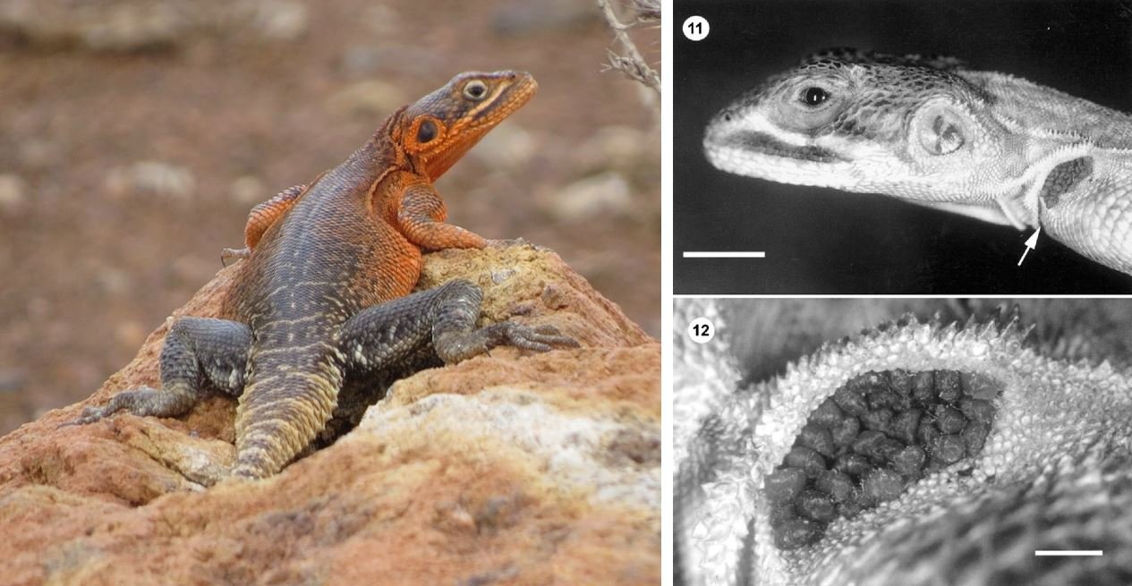
But are the pockets really ‘mite pockets’? You should have noticed so far a note of scepticism about Arnold’s ‘damage limitation hypothesis’. A follow-up to Arnold (1986) was published by Aaron Bauer, Anthony Russell and Norman Dollahon in 1990 following their analysis of hindlimb pockets in the New Caledonian gecko Rhacodactylus (Bauer et al. 1990).

Caption: a left, Gargoyle gecko Rhacodactylus auriculatus in captivity, the species studied by Bauer et al. (1990) in their test of Arnold’s hypothesis. At right, the ventral surface of the hindlimb of R. auriculatus, the popliteal fold and its entrance marked with an arrow. Images: Generish, CC BY-SA 4.0 (original here); Bauer et al. (1990).
And they concluded that the pockets weren’t anything to do with co-evolution and parasites, but a byproduct of the way lizard skin grows, folds or has evolved, the pockets – in cases – being related to use of the skin in crypsis and/or locomotion (Bauer et al. 1990). The mites, according to this view, are therefore taking advantage of sites of vulnerability on the lizards, and indeed damage caused to lizards by mites carries the same costs whether it occurs within the pockets or elsewhere on the body; it isn’t ‘limited’ as it should be according to Arnold’s hypothesis (Bauer et al. 1990).
An exchange between both sets of authors followed (Arnold 1993, Bauer et al. 1993), the ultimate conclusion (as seen by an outsider) being that the pockets are “neutral or mildly maladaptive “phylogenetic baggage”” (Bauer et al. 1993, p. 868). This was always my take on the phenomenon all along: that the pockets originated for reasons unrelated to co-evolution with a parasite, and that demonstrating lizard/mite co-evolution was a big ask, and a task that hadn’t been fulfilled. The similarity that the pockets have with various prominently marked or pigmented skin folds and flaps suggests, I think, that at least some of them are elaborations of structures that have roles in signalling or crypsis, and thus any use of them by parasites is incidental.

Caption: this is Flame the Central bearded dragon Pogona vitticeps, and like many lizards she has skin folds on the neck and shoulder region that are associated with distinctive ornamentation and markings. This suggests that the evolution of pockets (in the nuchal region at least) is an exaptation, and that the structures started out as being associated with signalling and display. Image: Darren Naish.
Mite pockets in the 21st century. 1993 is a long time along now. As I said at the start of this article, the general impression you might get from the literature today is that Arnold’s hypothesis has been accepted. I don’t know if this is because researchers really have been convinced by his proposal, or whether they’re aware of his initial argument (Arnold 1986, Benton 1987) but not the follow-up. Publications on lizards that have reason to refer to the pockets tend to just term them ‘mite pockets’ without equivocation (e.g., Rocha et al. 2008, Mockett 2017, Carvalho et al. 2018), though not all authors have done this (Fajfer 2012).

Caption: male specimen of the Bolivian-Brazilian tropidurid Tropidurus chromatops showing the deep and complex nuchal pockets of this species, in this case full of mites. Note how the pockets are also associated with distinctive pigmentation. Image: Carvalho et al. (2018).
Has any actual work been done on the pockets since the Arnold vs Bauer exchange of the 80s and 90s? Yes, yes it has, though let’s start with an observational report more than targeted experimental work. Bertrand & Modrý (2004) reported the discovery of numerous scale mites packed under a skin fold on the neck of the endemic Kenyan Elmenteita rock agama Agama caudospinosa. This lizard lacks proper pockets, and yet here was a case where clustering scale mites were causing a fold to become an incipient pocket.
Though the authors didn’t say it, this discovery potentially weakens the hypothesis that pockets evolved as mite aggregation sites: it looks instead like mite clustering is opportunistic, and that any suitable flap or skin fold is exploited.
Caption: at left, Elmenteita rock agama Agama caudospinosa in the field. At right, a specimen reported by Bertrand & Modrý (2004) in which numerous scale mites were packed under one of the nuchal skin folds. Images: rjq, CC BY-NC 4.0 DEED (original here); Bertrand & Modrý (2004).
Now for the big 21st century news on mite pockets: yes it’s true, Jay Clark Reed completed an entire 2014 doctoral thesis at the University of Michigan on the pockets and their possible role and evolution, albeit only in phrynosomatids (fence lizards and kin). So far as I can tell, a technical publication reporting the results of this work has yet to appear, so I feel a bit guilty discussing its results and hope I’m ok to do this. Reed’s (2014) primary conclusion was that the pockets probably did co-evolve with parasitic mites, since the included phylogenetic analyses showed that phrynosomatids evolved larger and more complex pockets on moving into mite-rich habitats. Meanwhile, pockets were reduced and lost in groups that – over the course of their evolutionary history – moved away from mite-heavy areas (Reed 2014). Yeah yeah, correlation isn’t causation, but this is at least suggestive, and consistent with Arnold’s hypothesis.
Reed’s mate selection hypothesis. Recall the mention early on that males carry higher tick loads than females? It’s well known that animals vetting others as potential mates partly base their evaluations on parasite load. Reed (2014) hypothesized that the pockets might have enlarged such that the deleterious presence of mites could be concealed, and thus make a parasite-loaded male more appealing to a female. This would mean that the pockets evolved as dishonest signals…. the theoretical problem here being that cheating about your parasite load (if that load is deleterious and hence worth concealing) would be bad in the long run, since it would result in offspring that are less fit than those of your competitors: those offspring would then lose out via natural selection. Err, unless everyone is lying about their parasite load I guess.

Caption: Yarrow’s spiny lizard Sceloporus jarrovii, the species used in Reed’s studies on mate selection and mite occurrence. S. jarrovii is a phrynosomatid iguania endemic to Mexico and the southern USA. Image: Greg Schechter, CC BY 2.0 (original here).
Anyway, via a series of ingenious tests (some involving the addition of painted-on pretend mites), it was shown that females did not make their mate selections on the basis of visible parasite load (Reed 2014). Ironically, females in some tests preferred males with the highest mite loads, but this is likely because big males – who are the most desirable – have the highest loads simply as a consequence of size. The idea that mate selection might have driven pocket evolution does not, therefore, seem supported (Reed 2014).
And that basically brings us to where we are. While further research is needed, mite pockets might really be mite pockets after all, and I find it exciting that a number of high-level evolutionary phenomena – exaptation, convergence, co-evolution, sexual selection and dishonest signalling among them – have all been suggested to have a bearing on this surprisingly odd and complex bit of anatomy. In addition, the fact that lizards have evolved such structures in the first place activates the speculative part of my brain…

Caption: certain species within the world of the Squamozoic have evolved elaborate pockets that have a very specialized function. In the giant subterranean amphisbaenian Graboidus, deep slits along the posterior part of the body lead to partitioned internal cavities inhabited by juveniles. Here, they feed on nutritious fluid expressed from the walls of the cavities. Image: Darren Naish.
I’ve been thinking about writing an article on mite pockets for years, but it was the sharing of a picture and its associated comments by digital artist and creature designer Kate Pfeilshiefter that inspired me to write this entire overly long article. Thank you, Kate.
For previous Tet Zoo articles on squamates that have some connection to this one, see…
Tale of the Takydromus, September 2013
Racerunner Lizards of the World Unite, October 2014
What's With All These New Chameleon Names?, Part 1, February 2016
By the Horns of Trioceros, the Casque of Calumma, the Brood of Bradypodion--Chameleons, Part 2, February 2016
Palleon, Archaius, Kinyongia, Nadzikambia--The Last Chameleons, Part 3, March 2016
Refs - -
Arnold, E. N. 1986. Mite pockets of lizards, a possible means of reducing damage by ectoparasites. Biological Journal of the Linnean Society 29, 1-21.
Arnold, E. N. 1993. Comment – function of the mite pockets of lizards: an assessment of a recent attempted test. Canadian Journal of Zoology 71, 862-864
Bauer, A. M., Russell, A. P. & Dollahon, N. R. 1990. Skin folds in the gekkonid lizard genus Rhacodactylus: a natural test of the damage limitation hypothesis of mite pocket function. Canadian Journal of Zoology 68, 1196-1201.
Bauer, A. M., Russell, A. P. & Dollahon, N. R. 1993. Function of the mite pockets of lizards: a reply to E.N. Arnold. Canadian Journal of Zoology 71, 865-868.
Benton, M. J. 1987. The mite pockets of lizards. Nature 325, 391-392.
Bertrand, M. & Modrý, D. 2004. The role of mite pocket-like structures on Agama caudospinosa (Agamidae) infested by Pterygosoma livingstonei sp. n. (Acari: Prostigmata: Pterygosomatidae). Folia Parasitologica 51, 61-66.
Carvalho, A. L. G., Rivas, L. R., Céspedes, R. & Rodrigues, M. T. 2018. A new collared lizard (Tropidurus: Tropiduridae) endemic to the Western Bolivian Andes and its implications for seasonally dry tropical forests. American Museum Novitates 3896, 1-54.
Curtis J. L. & Baird, T. A. 2008. Within-population variation in free-living adult and ectoparasitic larval trombiculid mites on collared lizards. Herpetologica 64, 189-199
Fajfer, M. 2012. Acari (Chelicerata) – parasites of reptiles. Acarina 20, 108-129.
Loveridge, A. 1925. A mite pocket in the gecko, Gymnodactylus lawderanus Stoliczka. Proceedings of the Zoological Society of London 1925, 1431.
McCoy, E. D., Styga, J. M., Rizkalla, C. E. & Mushinsky, H. R. 2012. Time since fire affects ectoparasite prevalence on lizards in the Florida scrub ecosystem. Fire Ecology 8, 32-40.
Mockett, S. 2017. A review of the parasitic mites of New Zealand skinks and geckos with new host records. New Zealand Journal of Zoology 44, 39-48.
Okabe, K. & Makino, S. 2008. Parasitic mites as part-time bodyguards of a host wasp. Proceedings of the Royal Society B 275, 2293-2297.
Reed, J. C. 2014. Analysis of the Function and Evolution of Mite Pockets in Lizards. University of Michigan Dissertation.
Rocha, C. F. D., Cunha-Barros, M., Menezes, V. A., Fontes, A. F., Vrcibradic, D. & Van Sluys, M. 2008. Patterns of infestation by the trombiculid mite Eutrombicula alfreddugesi in four sympatric lizard species (genus Tropidurus) in northeastern Brazil. Parasite 15, 131-136.
Wagner, P., Ineich, I., Leaché, A. D., Wilms, T. M., Trape, S., Böhme, W. & Schmitz, A. 2009. Studies on African Agama VI. Taxonomic status of the West African Agama (Sauria: Agamidae) with prominent tail crests: Agama boulengeri Lataste 1886, Agama insularis Chabanaud, 1918 and Agama cristata Mocquard, 1905. Bonner Zoologische Beiträge 56, 239-253.


